Vitamins, Minerals And Your Thyroid-What Your Doctor May Not Know
Shortly after hanging my Trichology ‘shingle’ I decided to specialise in female hair loss issues. I’d discovered early – contrary to general opinion – female hair loss is quite complex in what both influences and impels it.
Although males can (and do) experience different forms of alopecia, overwhelmingly the most commonly seen is Male Androgenic Alopecia – male ‘pattern’ balding. When a male has the genetics to exhibit this, it’s as much a natural part of post-pubertal secondary sex characteristics as facial whiskers, deepening voice, muscle bulk, and body hair.
By contrast thinning scalp hair in women is almost always an indication of internal dysfunction; a collapsing of body homeostasis to the point where hair growth can no longer be supported.
Iron, Vitamin D + Iodine (in that order) are considered three of the most important nutrients for optimal metabolic functioning. By virtue of a woman’s ‘femaleness’ she is more ‘at risk’ to be deficient in these nutrients than are males.
As a NON-essential skin appendage (in nutrient-metabolic-hormonal terms) hair is regularly the first tissue to have these supports withdrawn when body levels are becoming depleted. Hair shedding or a gradual thinning of scalp hair density or the activation of an autoimmune condition is often the initial symptom of internal disturbance or deficiency.
From menarche* to menopause it’s reasonable to assert most menstruating females will have some degree of iron deficiency at times in their life. Very few functions of the body are activated without sufficient iron to ‘furnace’ them.
Iron storage (termed ferritin) is considered the true indicator of iron status – with an accepted reference range of 20-300ug/L. To aspire to a ‘target’ level about mid-range – i.e.: 120-150ug/L** – could not be considered unrealistic given the importance of iron in the body (Lee: 2007).
The significance of reaching and maintaining this target level was the research of
Dr. John Lee – Australia’s most prolific thyroid researcher. Insufficient iron restricts cell mitochondria production from which Adenosine Tri-phosphate (ATP) – ‘cellular energy’ is created. Our metabolic activity and Phase II liver detoxification pathways are ATP dependant.
Regrettably some conformist practitioners still claim a ferritin of 21ug/L is within range and therefore ‘normal’! Just two points below (19ug/L) suggests ‘depleted iron stores’. To take this point further are they proposing a woman with a ferritin of 21ug/L (one point within range) will experience the equal energy and metabolic drive as another whose ferritin is 299ug/L (again one point within range)?
I also reject the claim of those traditionalists who say it’s impossible to achieve a 150ug/L ferritin in a pre-menopausal woman.
In terms of metabolic importance, Iodine is deemed the next most essential (trace) nutrient after iron. Simply put: Iodine deficiency = compromised thyroid hormone production.
Testing Iodine levels is a simple urinary ‘spot-screen’, but is seldom routinely assessed. Low Iodine results in an under-functioning thyroid. There is also a studied correlation between Iodine deficiency and reduced IQ in children, and breast disease in women.
At the time of writing – Australian Professor Creswell Eastman from the Council of Control (Iodine Deficiency Disorders) – is urging food manufacturers to again add Iodine to their products. His statement arises from a recent national study which found almost half of all children of primary school age show Iodine deficiency. A urinary Iodine test is NOT presently claimable under Australian Medicare.
The NHMRC recommends supplementation of 300-600mcg/day of Iodine to ensure women who are pregnant, breastfeeding or considering pregnancy have adequate Iodine status. Kelp (seaweed) supplements OR kelp-based products should NOT be taken as they may contain varying levels of Iodine AND be contaminated with heavy metals such as Mercury.
Research confirms Vitamin D*** is one of the three most important nutrients to health. Vitamin D is essential for the active absorption, utilisation + regulation of Calcium + Phosphorus within the body; it’s also vital for optimal thyroid functioning. Vitamin D is obviously ‘seasonal’; the reason people are often deficient at winter’s end.
Reference ranges (NEJM 357:3 July19th 2007) are 50-200 nmol/L. Levels less than 75nmol/L is considered Vitamin D insufficiency, whilst levels less than 50nmol/L are deemed ‘deficient’. Levels around 75nmol/L are essential to prevent Calcium loss from the body, and greater than 100nmol/L (revised ‘target’ level is 120nmol/L) for optimal thyroid functioning.
A 2009 study found women with Vitamin D levels greater than 85nmol/L had a 50% LOWER RISK of being diagnosed with breast cancer than those women with levels less than 60nmol/L (Rejnmark:2009).
Vitamin D deficiency has an adverse effect on hair growth due to its influence on thyroid-adrenal function. There are Vitamin D receptors in the scalp; Vitamin D is essential for hair follicle maturation (maturity). To positively influence any rT3, autoimmune issues or help stabilise blood sugar levels your Vitamin D must be maintained at 120-150nmol/L.
A Vitamin D deficiency can also trigger an autoimmune reaction in pre-disposed people as deficiency disorientates the immune system; attacking susceptible tissues such as the skin + the thyroid gland.
Progressive medical researchers**** speculate that just by annually testing and maintaining a woman’s Vitamin D and Iodine at respective ‘target’ levels, the rate of breast disease/malignancy could be decreased by as much as 25%..! They also propose the body requires less demand for Thyroid and Cortisol hormone when Iodine + Vitamin D levels are optimised – allowing the body to operate more efficiently.
Zinc is held to be implicated in at least 150 enzymatic actions within the body. Its main contributions to thyroid homeostasis are:
The synthesis of Thyrotropin Releasing Hormone (TRH) – produced by the Hypothalamus to stimulate production of Thyroid Stimulating Hormone (TSH) – also known as Thyrotrophin.
A crucial catalyst in the binding and activation of the active thyroid hormone Triiodothyronine (T3) to receptors on the cell nucleus.
Zinc deficiency is thought to contribute to poor thyroid hormone conversion – and deficiency diminishes healthy genetic expression of thyroid hormone.
A refractory zinc deficiency may result from inadequate protein availability (Baratosy: 2006). Amino acid (Tyrosine) derived from protein is a foundation of thyroid hormone production.
Reviewing copper (Cu) levels is particularly crucial. Low copper is said to inhibit thyroid gland hormone production, whilst elevated copper obstructs cell receptor interaction with thyroid hormone.
A deficiency of copper hinders the deployment of iron by the red blood cells, resulting in the iron being accumulated (and unavailable) within the organs of the body. Because this stored iron cannot be utilised whilst the copper deficiency persists, symptoms of iron deficiency may present – despite an actual iron sufficiency.
A refractory low-range or copper deficiency can be caused by Vitamin D deficiency (low Cu + Vitamin D go together).
If testing reveals BOTH zinc and copper to be deficient – this usually indicates malabsorption. Zinc and copper directly compete for absorption at the gut interface, so if one is elevated the other is habitually low or deficient.
An elevated copper level and Sex Hormone Binding Globulin (SHBG) is regularly seen in females using certain types of oral contraceptive. This is largely due to the additional (synthetic) oestrogen found in contraceptives and hormone replacement therapy. Oestrogen gives rise to copper retention – and vice versa – ultimately leading to zinc and other nutrient depletion, and oestrogen dominance.
SHBG is the ‘carrier’ protein for 70% of circulating, ‘bound’ (inactive) Testosterone (TT) and Oestrogen – and is produced in the liver. Other origins for elevated SHBG are:
Pregnancy
Hyperthyroidism
Cirrhosis of the liver
Medication such as Phenytoin Sodium (Dilantin) that induce hepatic enzyme induction
Once copper is in excess and too dominant in relation to zinc, it can exert what Baratosy (2005) describes as an ‘anti-nutrient’ – or toxic metal influence. High copper levels restrict the absorption and utilisation of zinc (particularly), iron, magnesium, Vitamins B3, 5, and 6, Vitamins C and E, and certain trace elements.
Sex Hormone Binding Globulin (SHBG) is produced in the liver, and is the carrier vitamins protein for (amongst other hormones) 70% of the circulating but ‘bound’ (inactive) testosterone and oestrogen. Elevated SHBG levels may result in symptoms of testosterone and oestrogen deficiency. A raised SHBG will also cause symptoms of low thyroid function because SHBG partly binds + inactivates the thyroid hormone T4.
In the long line of essential nutrients for optimal thyroid function, the importance of Selenium is only shaded by Iron and Iodine. Several thyroid enzymes are Selenium-dependant to the creation of thyroid hormone. Unlike copper and zinc, Selenium and Iodine are agonists to each other – with optimal levels of both (in balance) essential for a healthy thyroid gland. Selenium also has an integral role in anti-oxidant and immunity defence mechanisms.
There remain some differing opinions on the most reliable form of Selenium testing. Some advocate blood serum; others support hair mineral analysis (HTMA) – still others suggest toe nail clippings.
The B-vitamins are essential co-enzymes to maintaining mitochondrial ATP production. Compromised mitochondrial function leads to low metabolic (thyroid) activity. Thiamine (Vitamin B1), B12, Vitamin D and folic acid are synergistic to copper. Supplementing these nutrients where required helps restore body copper balance. Vitamin D metabolism is enhanced by copper. Adequate Vitamin D levels (>100nmol/L) are essential for optimal T3 receptor expression (Lee: 2007).
The Thyroid Hormones:
It’s not my intention to detail or even outline the anatomy and physiology of the thyroid-related endocrine system and the hormones involved. There are many excellent thyroid texts written by better educated and more qualified folk than me. I simply wish to convey to the lay reader what thyroid hormones they might request tested – and why:
Thyroid Stimulating Hormone (TSH): produced by the (anterior) Pituitary Gland – TSH regulates thyroid hormone production from the thyroid gland. TSH has long been regarded as the most reliable and sensitive indicator of thyroid function, however its limitations are these:
TSH does not reflect low metabolic activity; cell mitochondrial energy output and the necessary nutrients to furnace the body.
TSH does not reflect sufficient and quality conversion of the inactive thyroid hormone Thyroxine (T4) to the active, cell-influencing Triiodothyronine (T3).
TSH does not reflect deficiency of any of the numerous nutrients crucial to T4 – T3 synthesis, conversion, and activation.
TSH does not reflect T3 interaction with its mitochondrial or DNA receptors within the cell itself. If this interface fails – T3 cannot influence cell activity in any meaningful way.
TSH does not reflect elevated Reverse Triiodothyronine (rT3)***** levels which interfere with T4 – T3 conversion and T3’s activation of its intra-cell receptors.
TSH does not immediately reflect increasing thyroid antibodies in autoimmune thyroiditis.
Difficulties with any of the above has been termed ‘Euthyroid Sick Syndrome’ – patient’s exhibit symptoms of an under functioning thyroid but their TSH and T4 results are “normal”.
Thyroxine (T4): T4 is secreted by the thyroid gland in response to hypothalamic-pituitary stimulation (TRH/TSH). This secreted T4 then circulates in the blood – bound to a carrier protein – until synthesised (in the liver and kidneys) to T3. T4 possesses no interfacing receptors of its own, but is the inactive precursor of T3.
Triiodothyronine (T3): although some T3 is produced by the thyroid gland, greater than 80% results from T4 conversion. T3 is our active thyroid hormone which profoundly regulates body metabolism.
Reverse Triiodothyronine (rT3): rT3 is an adapted non-active form of Triiodothyronine (de-activated T4). In times of protracted physiological and emotional stress or illness, elevated Cortisol or Copper or heavy metal toxicity – T4’s normal conversion to T3 is corrupted – and rT3 results. Lee (2005) found forty percent of the synthetic thyroid hormone replacement Thyroxine sodium (Oroxine/Thyroxine etc) is altered to rT3.
In healthy, minimally-stressed people rT3 is quickly purged from the body. When rT3 levels are allowed to become excessive, they inhibit and distort T4 – T3 conversion – thus producing further rT3.
Elevating levels of rT3 is a ‘biological hibernation signal’ – shutting the body down to “await better times” (Van Zanden: 2012). Elevated rT3 levels are commonly detected in Chronic Fatigue and Fibromyalgia sufferers. Arem (1999) proposes these two debilitating illnesses are manifestations of thyroid dysfunction. A characteristic of ‘Wilson’s Thyroid Syndrome’ is patients’ exhibit high rT3 levels because T4 is continually corrupted to rT3 at the expense of T3.
Reverse T3 disrupts thyroid homeostasis by inhibiting the production and function of T3. rT3 binds to – but does not activate – T3 intra-cell receptors; effectively blocking T3 interface and activation.
Dr. John Lee was the first practitioner to facilitate the testing of rT3 in Australia.
Thyroid antibodies: thyroid antibodies are detectable indicators within the circulatory system that our immunity is primed against our thyroid gland. The presence of thyroid antibodies is sometimes discounted because a percentage of the population shows low levels of antibodies without any discernable thyroid disease.
Elevated levels typically signify autoimmune thyroiditis – ‘Hashimotos’ if the patient exhibits an under active thyroid state, and ‘Graves’ Disease’ if their symptoms/pathology suggest the thyroid is over active.
The usual thyroid antibodies tested in Australia are:
Thyroglobulin Antibodies
Thyroid Peroxidase Antibodies (TPO Ab) – the more sensitive test.
Researchers suggest a strong association between autoimmune thyroiditis and Coeliac Disease. Patients exhibiting both conditions were able to eliminate thyroid antibodies by adopting a Gluten-free diet (Baratosy: 2005). It’s believed BOTH Thyroid antibodies and Gluten antibodies originate from the gut and tend to go ‘hand-in-hand’. Most people exhibiting elevated thyroid antibodies have a ‘genetic marker’ for Gluten sensitivity when genetically-profiled (Cooper: 2011).
An Italian study of female nursing home geriatrics with hypothyroidism found that by eliminating gluten from the diet, the hypothyroid symptoms in these patients greatly diminished or disappeared.
The crucial roles sex and steroid hormones play in thyroid homeostasis – particularly Cortisol, Progesterone, and DHEA – have not been discussed here. Suffice to say the thyroid-adrenal relationship is mutually dependant, and a Saliva Hormone Assay of these and other relevant hormones is an integral part of the complete investigative process.
When treating thyroid-metabolic issues it’s essential to ‘balance’ the three key receptors: T3, D3 (Vitamin D) and CC (Cortisol). Medication/supplementation requirements depend on the interaction of these nuclear receptors – with T3 being the most important (Van Zanden: 2011).
It’s now known Vitamin D modifies thyroid receptors to a greater degree than previously realized, and D3 can drastically alter thyroid blood tests up or down. Of most concern to Practitioners are those patients who do not need much vitamin D – and if given too much – a hyperthyroid state may be induced (Van Zanden: 2011).
Toxic heavy metals – principally Lead, Mercury, Cadmium, Aluminum and Arsenic block the function of Vitamins and Minerals necessary for thyroid homeostasis. Where patients relate long-standing illness, toxic heavy metals should be an early assessment priority. Accurate and convenient testing is achieved by HTMA.
The thyroid hormone cascade is incredibly involved and complex. Vitamins, minerals, amino acids, trace elements, essential fatty acids (DHA/EPA), sex and steroid hormones, as well as the immune system must all be adequately available – and harmonious to each other – for T3 to accomplish its task. If any one of these vital components are lacking the process will stall – and optimal body functioning diminished.
In all this – hair is the expendable extravagance; usually the first tissue to suffer a withdrawal of metabolic and nutrient support.
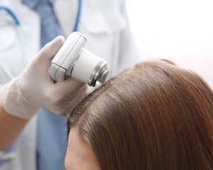
Scalp hair presentation:
In any of these nutritional or metabolic disturbances, scalp hair appearance and ‘feel’ often varies between individuals. Scalp hair may be lost from all over (diffuse); in some women a dual picture of female ‘pattern’ thinning with an underlying diffuse hair loss will be evident. The hair may feel observably dry or oily with thin, fragile hair shafts.
An anomaly of Zinc deficiency is dry, fragile/brittle hair BUT with an accompanying greasy/oily scalp. Other deficiency signs include poor wound healing, ‘white spotting’ on fingernails, lethargy, easy bruising and dry, scaly facial acne if the Zinc deficiency becomes severe.
Zinc is essential to optimal hair growth, and at physiological doses (50mgs/day) Zinc reduces Dihydrotestosterone (DHT) activity – the up-converted male hormone that influences androgenic hair loss. Zinc should only be taken for about 3-4 months at any one time as it will oppose copper & iron absorption & utilisation.
Thinning scalp hair resulting from severe thyroid disturbance frequently presents as ‘pattern’ thinning descending down to ear level, with an underlying diffuse hair thinning. The hair itself has a fine ‘cotton wool’ feel to it which I term ‘thyroid hair’. If autoimmune activity is involved (i.e.: autoimmune thyroiditis or accompanying autoimmune condition) – loss of eyebrows (partially or fully); decreased body hair and a thinning or loss of eyelashes will also be apparent.
It should now be appreciated that “gimmicky” single treatments such as laser combs, commercial hair loss programs etc can do nothing to influence nutritional, metabolic or hormonal disturbance. These areas must be individually tested for –and reviewed and treated as part of the total picture.
About the Author: Tony Pearce WTS is a Specialist Trichologist of female hair loss + scalp problems. He is a Member of the World Trichology Society (WTS), Vitamin D Council (USA) and Associate Member of the Australasian College of Nutritional + Environmental Medicine (ACNEM). Copyright Anthony Pearce 2008 (revised May 2013)
*The onset of menstruation in a young female at puberty.
** A Post-menopausal woman should be around 100ug/L
*** Read more in-depth information on Vitamin D at www.hairlossclinic.com.au (Vitamin D – the re-discovered key to illness prevention). **** Dr. John Lee and ACNEM Doctors/Natural Medicine Practitioners
*****See Post (‘When the Body Shoots Blanks’) at my Blog – www.hairlossclinic.com.au which explains rT3 in plainer terms for non-medical people. The consequences of elevated rT3 is UNDER-active thyroid-like symptoms; tiredness, thinning scalp hair density, weight gain, mood disturbance.